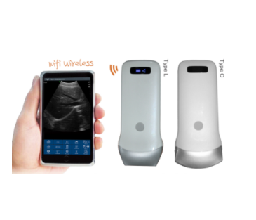
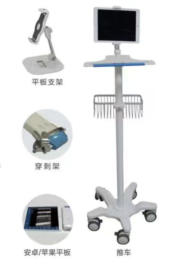
C6/C7 Series Technical Specifications
Product Versions
1. C6: wireless mini B-mode ultrasound scanner.
2. C7: wireless mini color ultrasound scanner.
3. Detailed imaging modes and functions depend on the selected configuration.
Scanning Method
Electronic array scanning.
Probe Frequency
3.5MHz and 5.0MHz.
Scanning Depth
90mm, 160mm, 220mm and 305mm, adjustable.
Imaging Modes
1. C6 supports B and B/M modes.
2. C7 color imaging modes depend on the actual configuration.
Image Adjustment
1. Gain adjustment.
2. Focus adjustment.
3. Pulse-inversion harmonic imaging.
4. Noise reduction.
Puncture Assistance
1. In-plane puncture-guidance line.
2. Out-of-plane puncture guidance.
3. Automatic vessel measurement.
4. Needle-tip enhancement.
Battery and Charging
1. Battery operating time: approximately 3 hours.
2. Wireless charging.
3. USB charging.
4. Operating time can be extended with a wireless power bank.
Measurement Functions
1. Distance.
2. Area.
3. Circumference.
4. Heart rate.
5. Obstetric measurements.
Cine-Loop Playback
Manual and automatic cine-loop playback with 100, 200, 500 or 1000 frames.
File Storage
Supports JPG, AVI and DICOM formats.
Wireless Connection
1. Wi-Fi connection to smartphones, tablets and Windows devices.
2. Wi-Fi standard: 802.11n.
3. Frequency band: 2.4GHz.
Image Frame Rate
Approximately 14 frames per second.
Software Compatibility
Apple iOS, Android and Windows.
Dimensions and Weight
1. Dimensions: 156mm × 60mm × 20mm.
2. Weight: approximately 250g.
Product Name: C6/C7 Wireless Mini Ultrasound Scanner
Product Overview
The C6/C7 series is a wireless mini ultrasound system that connects to smartphones, tablets and Windows devices through Wi-Fi.
C6 is the black-and-white B-mode version, while C7 is the color ultrasound version. The compact,
wireless and waterproof design makes the system easy to carry, clean and disinfect.
Main Features
1. C6 B-mode and C7 color ultrasound versions.
2. Smartphone, tablet and Windows compatibility.
3. Cable-free operation.
4. Compact and lightweight design.
5. Waterproof housing for convenient cleaning and disinfection.
6. Wireless and USB charging.
7. Puncture guidance.
8. Automatic vessel measurement.
9. Needle-tip enhancement.
10. Image storage and cine-loop playback.
11. Remote image sharing and consultation.
Basic Operating Procedure
1. Check the battery level of the wireless probe and display device.
2. Install the designated application on a smartphone, tablet or Windows device.
3. Turn on the C6 or C7 probe.
4. Connect to the probe Wi-Fi signal according to the software instructions.
5. Select the correct model and examination mode.
6. Apply ultrasound gel to the probe or examination area.
7. Position the probe and observe the real-time image.
8. Adjust gain, depth, focus, harmonic imaging and noise reduction as required.
9. Activate the appropriate puncture-guidance function when needed.
10. Save the required images, videos or DICOM files.
11. Turn off the application and probe after the examination.
12. Clean and disinfect the device according to the product instructions.
Precautions
1. The device should be operated by trained ultrasound professionals.
2. C6 and C7 have different imaging functions, so the correct model should be selected.
3. Strict aseptic procedures must be followed during puncture-guided operations.
4. Make sure that disposable protective covers do not significantly affect image quality.
5. The intended use is subject to the product registration certificate and local regulations.
C6/C7 Wireless Mini Ultrasound Images
The C6/C7 series can display multiple clinical ultrasound images, including:
1. Abdominal ultrasound images.
2. Obstetric ultrasound images.
3. Superficial-tissue images.
4. Vascular ultrasound images.
5. B/M-mode images.
6. Puncture-guidance images.
7. C7 color ultrasound images, depending on the actual configuration.
The images shown on this page are provided only to demonstrate the imaging functions of the products.
Examinations and diagnosis should be performed by trained professionals.