(0086) 755 2882 8917

Xindavet

sunway

C60
Others
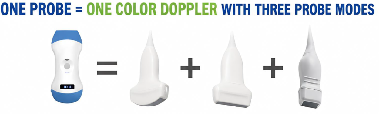
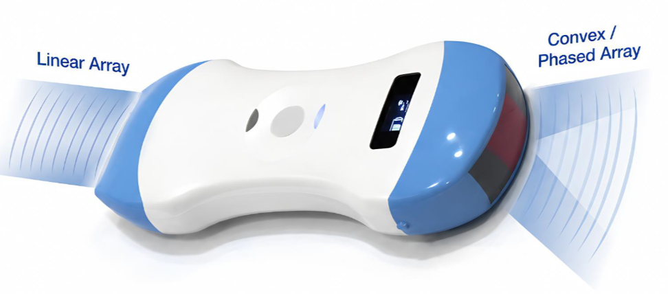
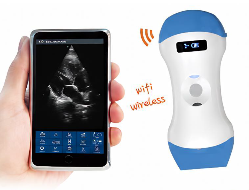
C60 is a 3-in-1 wireless handheld color Doppler ultrasound scanner integrating convex, linear and phased-array scanning modes in a single probe. The convex mode is suitable for abdominal and obstetric examinations, the linear mode for superficial tissue and vascular examinations, and the phased-array mode for cardiac examinations. The system supports B, B/M, Color, PW and PDI imaging modes and connects wirelessly to smartphones, tablets and Windows devices through Wi-Fi. Its compact and lightweight design makes it easy to carry, while the cable-free probe provides greater freedom of movement. The waterproof housing also makes cleaning and disinfection more convenient. C60 supports wireless charging, image and video storage, cine-loop playback, puncture guidance and automatic vessel measurement. Images can also be conveniently shared for remote consultation using the communication functions of a smartphone or tablet.
  • SUPPORT
  • SPECIFICATIONS
  • OPTION
  • GALLERY

Technical Specifications


Scanning Method


Electronic array scanning.


Elements and Channels


1. Number of elements: 128.

2. Number of channels: 32.


Probe Modes and Frequencies


1. Convex frequency: 3.2MHz and 5.0MHz.

2. Phased-array frequency: 2.5MHz and 5.0MHz.

3. Linear frequency: 7.5MHz and 10MHz.

4. One probe integrates both convex and linear transducer heads.

5. The convex mode can be switched to phased-array mode through the software.


Scanning Depth


1. Convex: 90mm, 160mm, 220mm and 305mm.

2. Phased array: 90mm, 120mm, 140mm and 160mm.

3. Linear: 20mm, 40mm, 60mm and 80mm.

4. Adjustable scanning depth.


Scanning Angle and Width


1. Convex scanning angle: 45 degrees.

2. Phased-array scanning angle: 80 degrees.

3. Linear scanning width: 40mm.


Imaging Modes


B, B/M, Color, PW and PDI.


Image Adjustment


Gain, focus, pulse-inversion harmonic imaging and noise reduction.


Puncture Assistance


1. In-plane puncture-guidance line.

2. Out-of-plane puncture guidance.

3. Automatic vessel measurement.


Battery and Charging


1. Battery operating time: approximately 3 hours.

2. Wireless charging.

3. The operating time can be extended with a compatible wireless power bank.


Image and Measurement Functions


1. Image frame rate: 18 frames per second.

2. Measurements: distance, area, circumference, heart rate and obstetric measurements.

3. Manual and automatic cine-loop playback.

4. Cine-loop frames: 100, 200, 500 or 1000.

5. Image and video formats: JPG, MP4, PNG and DCM.


Wireless Connection


1. Wi-Fi connection between the probe and display device.

2. Supports 802.11n.

3. Supports 2.4GHz and 5GHz dual-band Wi-Fi.

4. Transmission speed up to 450Mbps.


Software Compatibility


Apple iOS, Android and Windows.


Dimensions and Weight


1. Dimensions: 156mm x 60mm x 20mm.

2. Weight: approximately 230g.


Product Name: C60 3-in-1 Wireless Handheld Color Doppler Ultrasound Scanner


Product Overview


C60 is a wireless handheld color Doppler ultrasound system combining convex, linear and phased-array scanning modes in a single probe. It connects to smartphones, tablets or Windows devices through Wi-Fi and displays real-time ultrasound images without a conventional probe cable.


Main Applications


1. Convex mode: abdominal, obstetric and deep-tissue examinations.

2. Linear mode: vascular, superficial, musculoskeletal and puncture-guidance examinations.

3. Phased-array mode: cardiac and intercostal examinations.

4. Color Doppler mode: assists in visualizing blood flow.

5. PW mode: pulsed-wave Doppler spectrum observation.

6. PDI mode: low-velocity blood flow and perfusion imaging.


Main Features


1. Three scanning modes integrated into one probe.

2. Supports B, B/M, Color, PW and PDI.

3. Compact and lightweight for easy carrying.

4. Wireless operation without a probe cable.

5. Waterproof housing for convenient cleaning and disinfection.

6. Wireless charging.

7. Puncture assistance and automatic vessel measurement.

8. Supports JPG, PNG, MP4 and DCM formats.

9. Compatible with iOS, Android and Windows.

10. Convenient image sharing and remote consultation.


Basic Operating Procedure


1. Make sure the C60 probe and display device are sufficiently charged.

2. Install the designated application on a smartphone, tablet or Windows device.

3. Turn on the C60 probe.

4. Connect to the C60 Wi-Fi signal according to the software instructions.

5. Select convex, linear or phased-array mode according to the examination area.

6. Select B, B/M, Color, PW or PDI imaging mode.

7. Apply a suitable amount of ultrasound gel to the probe or examination area.

8. Position the probe and observe the real-time ultrasound image.

9. Adjust gain, depth, focus, harmonic imaging and noise reduction as required.

10. Activate the appropriate puncture-assistance function when performing a guided procedure.

11. Save the required images or videos after the examination.

12. Turn off the application and probe, then clean and disinfect the device according to the instructions.


Precautions


1. The device should be operated by personnel trained in professional ultrasound examination.

2. Select the correct scanning mode for the examination area.

3. Follow clinical aseptic procedures during puncture-guided operations.

4. Make sure that any disposable protective cover does not significantly affect image quality.

5. Clean and disinfect the device according to the product instructions.

6. Software interfaces and functions may vary depending on the version.

7. The intended use of the product is subject to its registration certificate and local regulations.


C60 3-in-1 Wireless Ultrasound Scan Images


C60 provides multiple clinical ultrasound imaging applications through a single probe, including:


1. Convex abdominal ultrasound images.

2. Convex obstetric ultrasound images.

3. Linear vascular ultrasound images.

4. Linear superficial-tissue images.

5. Linear musculoskeletal images.

6. Phased-array cardiac ultrasound images.

7. Color Doppler blood-flow images.

8. PW Doppler spectrum images.

9. PDI power Doppler images.

10. Puncture-guidance images.


The images shown on this page are provided only to demonstrate the imaging functions of the product. Examinations and image interpretation should be performed by trained medical professionals.


Transducers

One Probe